Método de detección de células senescentes
Nov 17, 2025
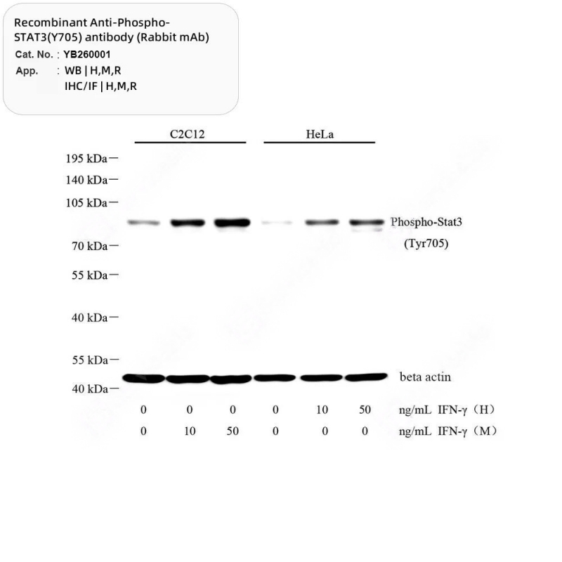
I. Panorama general de la senescencia celular La senescencia celular es un estado irreversible de detención del crecimiento al que entran las células normales tras un número limitado de divisiones, lo que actúa como mecanismo de protección para controlar el potencial proliferativo. Las células senescentes experimentan cambios en su morfología y actividad metabólica; por ejemplo, un aumento del volumen celular y una mayor regulación de la actividad de la β-galactosidasa asociada a la senescencia (SA-β-gal), que se convierte en un marcador de detección clásico.II. Método de detección de núcleos: tinción de SA-β-gal Principio: La β-galactosidasa en los lisosomas de las células senescentes es activa a un pH de 6,0 (a diferencia del pH normal de 4,0) y puede catalizar el sustrato X-Gal para producir un producto azul.Aplicación: El precipitado azul se observa mediante microscopía óptica. Aplicable a células, secciones congeladas y preparaciones completas de tejido.Productos recomendados:Fijador especial (Cat. n.° YB2225)Kit de tinción celular (cat. n.° YB2173)Kit de tinción de tejido completo (n.° de cat. YB2241, incluye solución aclaradora, permite seccionar después de la tinción de montaje completo)III. Marcadores de senescencia y recomendaciones de anticuerposSegún las pautas de Cell, los marcadores de senescencia se clasifican en varias clases; se recomienda su uso combinado para mejorar la especificidad:Marcadores de detención del ciclo celularp21 (Cat# YB265313 / YB260038): Un inhibidor de CDK corriente abajo de p53, clave para el arresto irreversible.p16INK4a (Cat# YB261143 / YB261605): El marcador de senescencia más específico, regulado positivamente de manera constante.Marcadores de proliferación negativosKi67 (Cat# YB221499 / YB260023): se utiliza para excluir células proliferantes y confirmar el arresto del ciclo.PCNA (Cat# YB22010 / YB23010): la expresión disminuida sugiere arresto, mientras que los niveles aumentados podrían indicar reparación del daño del ADN.Estructura nuclear y alteraciones de la cromatinaLámina B1 (Cat# YB221802 / YB261802): La pérdida de esta proteína de la lámina nuclear conduce a la pérdida de la integridad nuclear.HMGB1 (Cat# YB26103 / YB22103): Pérdida de localización nuclear y liberación al espacio extracelular, lo que impulsa la senescencia inflamatoria.γH2A.X (Cat# YB221841): Marcador de roturas de doble cadena de ADN (fosforilado en Ser139).Fenotipo secretor asociado a la senescencia (SASP)IL-6, IL-1β, IL-8, TNF-α (Cat# YB22117 / YB22113, etc.): citocinas proinflamatorias que dan forma a un microambiente inflamatorio crónico.p-STAT3 (Cat# YB260001): Molécula de señalización clave que activa SASP y la reprogramación metabólica.Metabolismo y marcadores de estrés oxidativo4-HNE (Cat# YB260073): Producto de peroxidación lipídica cuya acumulación impulsa la senescencia.MMP3 (Cat# YB26131): degrada la matriz extracelular y participa en la remodelación tisular.IV. Puntos clave del diseño experimentalUso de marcadores multiplex: Los marcadores individuales son propensos a falsos positivos. Es esencial una combinación que integre la tinción de SA-β-gal, proteínas de detención del ciclo celular (p16/p21), marcadores de proliferación negativos (Ki67), etc., para una evaluación completa.Referencia de validación in vivo: el texto proporciona una tabla de control positivo para la evaluación de la senescencia en tejidos de ratón (por ejemplo, expresión de p21 en hígado envejecido, reducción de Lamin B1 en tejido cerebral), que puede guiar la selección del modelo.ResumenLa detección de células senescentes requiere un cribado inicial mediante tinción de la actividad de SA-β-gal, seguido de una validación con marcadores multidimensionales, como proteínas reguladoras del ciclo celular, cambios estructurales nucleares y factores SASP. Al seleccionar anticuerpos, asegúrese de que los números de catálogo coincidan con el sistema experimental (humano, ratón, etc.) y el método de detección (WB, IHC/IF) para garantizar la fiabilidad de los resultados.


 RED SOPORTADA
Xml / política de privacidad
RED SOPORTADA
Xml / política de privacidad Español
 English
 Русский
 Español